
ANATOMY AND PHYSIOLOGY
Divers need a basic understanding of anatomy and physiology to cope with the heightened demand imposed on the body during underwater activities.
The Circulatory System
The heart is a hollow, muscular organ the size of a fist. It sits in the center of the chest just behind the breastbone and in a space between the lungs which is called the mediastinum. In man, the right and left halves of the heart normally have no direct connection.
Each half of the heart is divided into an upper chamber, the atrium, which receives blood from the veins, and a lower chamber, the ventricle, which pumps this blood into the arteries. The ventricles are more muscular chambers than the atria and do most of the work of pumping.
The diffusion of gases between blood and air occurs through the thin walls of microscopic blood vessels called capillaries. These surround the tiny air sacs in the lungs known as alveoli. Similar capillaries in the body tissues allow gas exchange between cells and blood.

Alveolar Gas Exchange
Alveolar Gas Exchange PCO2 (Partial Pressure of CO2) PO2 (Partial Pressure of O2)
PCO2 (Venous Blood) > PCO2 (Alveolar) Therefore CO2 passes from the blood to the Lungs.
PO2 (Venous Blood) < PO2 (Alveolar) Therefore O2 is taken up by blood from the lungs.

Cellular Gas Exchange
PCO2 (Cell) > PCO2 (Arterial) Therefore CO2 passes from the cell to the blood. PO2 (Arterial Blood) < PO2 (Cell) Therefore O2 is taken up by cells from the blood.
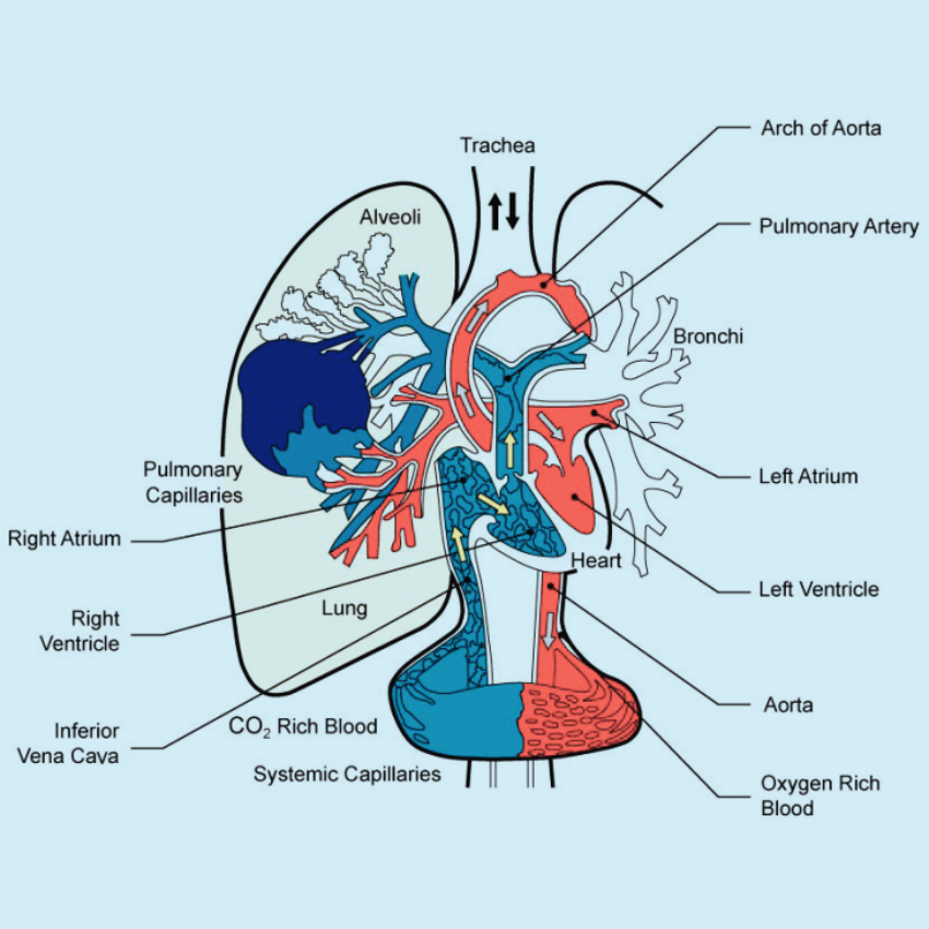
Oxygen-rich blood leaves the lungs and is carried to the left atrium of the heart by the pulmonary veins. The blood is then pumped into the left ventricle and from there, through a series of increasingly smaller arteries, to ‘beds’ of capillaries in the body tissues. Veins collect blood from these capillaries and join one another to form progressively larger vessels that empty back into the right atrium of the heart. From here blood is pumped into the right ventricle and then back to the lungs (see image).
The walls of arteries are rich in elastic and muscle fibers, which makes them strong and resilient to the high blood pressures produced by the ventricles. The muscles within the walls of arteries are arranged so that the diameter of the vessels and the distribution of blood within the body may be controlled. Veins need only thin walls because they carry blood at low pressure; they contain valves to ensure that blood flows in the right direction.
On each side of the heart, there is a valve between the atrium and ventricle and between the ventricle and its outflow artery. These non-return valves ensure that blood is pumped in the correct direction.
An average human body contains about 6 liters of blood. The blood contains billions of red cells to enable it to carry a sufficient amount of oxygen. These are microscopic disc-shaped cells, which are packed with hemoglobin, an iron-containing compound that binds loosely with oxygen. When blood is rich in oxygen the ox-hemoglobin gives it a bright red color; a low oxygen concentration results in a bluish tinge.
A rare inherited disorder known as ‘Sickle-Cell Disease’ is caused by abnormal hemoglobin: this results in the red cells assuming an irregular shape in conditions of low oxygen. Since these red cells are not very flexible, sickle-cell disease can result in tissue injury due to the blockage of small blood vessels. Therefore, individuals with this condition are not allowed to dive. A small amount of carbon dioxide can be carried dissolved in the blood as carbonic acid and the rest is carried by the red cells.
SUMMARY OF THE FUNCTION OF THE CIRCULATION
The function of blood circulation is to carry to the tissues of the body the substances they require and to remove from them their secretions and the waste products they produce in their activities. Blood enters the Pulmonary Capillaries, which surround the Alveoli in the lungs via the Pulmonary Artery having been pumped from the Right Atrium and then the Right Ventricle.

The blood takes on O2 and gives off CO2 and then passes to the Left Atrium via the Pulmonary Vein. The blood is then pumped via the Left Ventricle into the main Artery (The Aorta). The O2-rich Arterial Blood is then passed via the Arterial system to the body’s cells where the O2 is passed to the cells and the CO2 is removed. The now O2-depleted and CO2-rich blood (Venous Blood) is then returned to the Right Atrium of the Heart via the Vena Cava. On entry to the Right Atrium, the process is repeated.
The amount of blood in the body does not normally change very much. However, the rate at which it circulates depends greatly on the needs of tissues. The greater the rate at which oxygen is used up by a tissue, the greater the amount of blood, which is supplied to it. As the body’s demand for oxygen increases so the pulse rate and the volume of blood pumped by each beat of the heart increase accordingly. At rest, the heart normally beats roughly 70 times and pumps about 5 liters of blood per minute. During heavy exercise, the pulse of a fit young person can exceed 180 beats, and more than 20 liters of blood can be pumped each minute.
The blood pressure has to remain high enough to ensure an adequate supply of blood to the whole body but low enough to avoid bursting the more delicate blood vessels. The blood pressure is monitored by structures, which lie in the carotid arteries (which supply the head and neck) and in the aorta (the body’s main artery). Information from these is used by the brain to control blood pressure using hormones and the autonomic nerve supply to the heart and blood vessels.
Blood pressure is usually measured in millimeters of mercury (mmHg). This reaches a maximum when the heart is contracting (systolic pressure), and a minimum value between beats (diastolic pressure). In a fit young person at rest, the systolic pressure is about 120 mmHg and the diastolic pressure is about 80 mmHg. Both pressures are
usually measured and are written down as a fraction with the systolic above and diastolic below (e.g. 120/80). Both pressures increase considerably during exertion and excitement, but if they remain high at rest then there may be some abnormality. A certain amount of increase in blood pressure is natural with age because the arteries gradually lose some of their elasticity.
In an emergency, adrenaline is released by the adrenal glands into the blood in response to a nervous signal from the brain. This increases heart rate and blood pressure in readiness for intense activity.
The automatic control of blood pressure is occasionally upset by stress. A highly unpleasant emotion (for example that brought on by pain or the sight of a gory injury) may have this effect. When the control is upset, the blood pressure may fall to the point where insufficient blood reaches the brain. This can cause dizziness, weakness, nausea, and, eventually, loss of consciousness – fainting. This causes the victim to fall, which lowers the head and thus permits more blood to reach the brain. Thus, consciousness usually returns within a few minutes. Collapse caused by fainting can often be averted by lowering the head – e.g. sitting down with the head between the knees.
Fainting may also occur if someone stands still for a long time (especially in hot weather) or very suddenly stops strenuous exertion. This occurs because blood pools in the lower part of the body and does not flow back to the heart sufficiently rapidly to allow the maintenance of adequate blood pressure. Such fainting is uncommon while diving because water immersion supports the circulatory system so that pooling of blood does not occur. Lightheadedness may occasionally be experienced on leaving the water, particularly if the water was warm and there was heavy exertion during the dive. However, it is important to realize that collapse after diving may be caused by more serious disorders.
Shock is a serious condition, which is caused by a loss of blood or plasma from the circulation. Severe internal and external wounds and burns commonly cause shock. When the body is unable to maintain blood pressure, tissues are starved of oxygen, and waste products build up. This can be fatal and must be treated rapidly. The
pulse in someone with shock is usually weak and very rapid. Medical attention and fluid replacement are required urgently.
Conclusion:
The circulatory system plays a vital role in transporting essential substances and removing waste products. It ensures tissue oxygenation, nutrient delivery, and waste removal for overall body function and health. In addition, the circulatory system maintains blood pressure, regulates blood flow, and adapts to the body’s demands, allowing for optimal functioning and adaption during various activities and conditions